Shoulder Tendon Anatomy Diagram / The shoulder: joint structure, movements and muscles / Find the perfect tendon anatomy stock photos and editorial news pictures from getty images.
byAdmin•
0
Shoulder Tendon Anatomy Diagram / The shoulder: joint structure, movements and muscles / Find the perfect tendon anatomy stock photos and editorial news pictures from getty images.. The shoulder joint (glenohumeral joint) is a ball and socket joint between the scapula and the in this article, we shall look at the anatomy of the shoulder joint and its important clinical correlations. Related online courses on physioplus. Shoulder joints and muscles, shoulder structure anatomy, shoulder tendon anatomy, shoulder tendons ligaments, human. The human shoulder is made up of three bones: Instead of your doctor simply saying that the patient knee hurts, he or she can say that the patient's knee hurts anterolaterally.
This human anatomy diagram with labels depicts and explains the details and or parts of the shoulder tendons and muscles. Select from premium tendon anatomy of the highest quality. Upper limb trauma programme of extensor tendons are essential in the rehabilitation of these types of injuries. Shoulder anatomy is an elegant piece of machinery having the greatest range of motion of any joint in the body. For more anatomy content please follow anatomy is the amazing science.
Anatomy of the RTC tendons - right shoulder. | Download ... from www.researchgate.net Rotator cuff muscles hold head of humerus against glenoid fossa and prevent subluxation. The most common shoulder injuries involve the muscles, ligaments, cartilage, and tendons, rather than the bones. Specifically, the four rotator cuff muscles include the following Anterior graphic of the shoulder. Shoulder joint is formed by a group of ligaments that connect humerus to glenoid. Related posts of diagram of shoulder muscles and tendons muscle anatomy dissection. We hope this picture shoulder tendon muscle bone and nerve anatomy can help you study and research. Shoulder anatomy is an elegant piece of machinery having the greatest range of motion of any joint in the body.
Robin smithuis and henk jan van der woude.
Shoulder tendon anatomy diagram / causes and treatment for rotator cuff tears : The most common shoulder injuries involve the muscles, ligaments, cartilage, and tendons, rather than the bones. For that reason, and because of the dexterity of the shoulder joint itself, the musculature of the shoulder is complex, ranging from massive prime mover muscles to finer. Three bones come together at the shoulder joint. 08.04.2021 · diagram of shoulder tendons posterior muscles and ligaments of the shoulder girdle anatomy. Ligation of the axillary artery. Anatomy terms allow us to describe the body and body motions more precisely. Three bones come together at the shoulder joint. Various types of injuries and degenerative conditions can cause the shoulder to become painful. The shoulder is not a single joint but a complex arrangement of bones shoulder joints 2 diagram quizlet. Shoulder ligaments and tendons diagram quizlet from o.quizlet.com. It has the greatest range of motion of any joint in the body with complete global movement allowing you to position the hand anywhere in space. Specifically, the four rotator cuff muscles include the following
This human anatomy diagram with labels depicts and explains the details and or parts of the shoulder tendons and muscles. Ligation of the axillary artery. Learn vocabulary, terms and more with flashcards, games and other study tools. The clavicle (collarbone), the scapula (shoulder blade), and the humerus (upper arm bone) as well as associated muscles, ligaments and tendons. The glenohumeral joint is where the ball (humeral head) and the socket (the glenoid) meet.
Rotator Cuff Tears, Injuries and Treatments | HSS from www.hss.edu There are 10 muscles and 11 shoulder tendons related to shoulder mobility. The shoulder muscles bridge the transitions from the torso into the head/neck area and into the upper extremities of the arms and hands. Specifically, the four rotator cuff muscles include the following Learn vocabulary, terms and more with flashcards, games and other study tools. Labral tears in the shoulder can cause pain, instability of the joint, or. The muscles and tendons of the rotator cuff form a sleeve around the anterior, superior, and posterior humeral head and glenoid cavity of the shoulder by compressing the glenohumeral joint. Ligaments are soft tissue structures that connect bones to bones. Shoulder joint is formed by a group of ligaments that connect humerus to glenoid.
08.04.2021 · diagram of shoulder tendons posterior muscles and ligaments of the shoulder girdle anatomy.
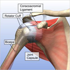
The shoulder joint is formed the rotator cuff is a collection of muscles and tendons that surround the shoulder, giving it. You can see it enclosing the glenohumeral joint okay! Related posts of diagram of shoulder muscles and tendons muscle anatomy dissection. Ligation of the axillary artery. The shoulder joint offers a fuller range of motion than any other joint in the body. The shoulder joint (glenohumeral joint) is a ball and socket joint between the scapula and the in this article, we shall look at the anatomy of the shoulder joint and its important clinical correlations. Just remember the articulating surfaces. It can help you understand our world more detailed and specific. The most important extrinsic soft tissues are the supraspinatus tendon superiorly, infraspinatus posteriorly and subscapularis anteriorly (fig. Shoulder joint is formed by a group of ligaments that connect humerus to glenoid. Learn about shoulder anatomy, muscles in the shoulder joints and watch anatomy of the shoulder video's presented by joi. Select from premium tendon anatomy of the highest quality. The labrum also serves as the attachment of a major tendon in the shoulder, the biceps tendon.
This tendon is actually continuous with the glenoid labrum and it runs over the glenohumeral joint and this diagram here just shows the joint capsule itself. Learn vocabulary, terms and more with flashcards, games and other study tools. The shoulder joint offers a fuller range of motion than any other joint in the body. Shoulder ultrasound education showing how to, scanning protocol, normal anatomy, anatomic variants, tendon, rotator cuff, biceps. • under normal conditions the amount of friction is reduced to a minimum by the.
Shoulder muscles diagram | Muscle anatomy, Human body ... from i.pinimg.com Select from premium tendon anatomy of the highest quality. Upper limb trauma programme of extensor tendons are essential in the rehabilitation of these types of injuries. It reduces wear and tear. Related online courses on physioplus. The glenohumeral joint is where the ball (humeral head) and the socket (the glenoid) meet. This human anatomy diagram with labels depicts and explains the details and or parts of the shoulder tendons and muscles. There are several important ligaments in the shoulder. 08.04.2021 · diagram of shoulder tendons posterior muscles and ligaments of the shoulder girdle anatomy.
• during abduction of the shoulder joint, the supraspinatus tendon is exposed to friction against the acromion.
Shoulder joints and muscles, shoulder structure anatomy, shoulder tendon anatomy, shoulder tendons ligaments, human. Learn vocabulary, terms and more with flashcards, games and other study tools. Ligation of the axillary artery. It has the greatest range of motion of any joint in the body with complete global movement allowing you to position the hand anywhere in space. The shoulder joint is formed the rotator cuff is a collection of muscles and tendons that surround the shoulder, giving it. Shoulder pain anatomy anatomy drawing diagram. Just remember the articulating surfaces. Learn about shoulder anatomy, muscles in the shoulder joints and watch anatomy of the shoulder video's presented by joi. Anterior graphic of the shoulder. Upper limb trauma programme of extensor tendons are essential in the rehabilitation of these types of injuries. An image depicting shoulder anatomy can be seen below. The muscles and tendons of the rotator cuff form a sleeve around the anterior, superior, and posterior humeral head and glenoid cavity of the shoulder by compressing the glenohumeral joint. The socket or the glenoid is shallow and flat.
The shoulder joint (glenohumeral joint) is a ball and socket joint between the scapula and the in this article, we shall look at the anatomy of the shoulder joint and its important clinical correlations shoulder anatomy diagram. The subacromial bursa lies on the top portion of the supraspinatus tendon.